Pleural Fluid Color Chart
Pleural Fluid Color Chart - Web in addition to its diagnostic value, pleural fluid analysis also has predictive value (ie, estimates of the likelihood of a clinical response to pleural fluid drainage) and. This is the space between the lining of the outside of the lungs (pleura). Web learn how to examine pleural fluid by protein, ldh, glucose, ph and other tests. Web interpreting pleural fluid results correctly requires an awareness of the possible aetiologies of a pleural effusion and an understanding of the reliability of the outcome of each. Pleural fluid color will be obviously abnormal (e.g., bilious or black). Under normal circumstances, a small amount of fluid is continuously produced and. Web the aims of this study were to describe the different appearances of pleural fluid during thoracentesis and their frequency in relation to diagnosis, and to evaluate the causes. An observant practitioner of thoracentesis cannot help but appreciate the similarity between pleural. 863 iu/dl) were statistically higher in bloody pleural. Web a pleural effusion is an abnormal fluid accumulation within the pleural space. An observant practitioner of thoracentesis cannot help but appreciate the similarity between pleural. Your provider has to get rid of this fluid with medicine or a. Web the number of red blood cells (220.5 × 10 3 /μl vs. 863 iu/dl) were statistically higher in bloody pleural. Find out the common causes and differential diagnosis of exudate and. Colour of pleural effusion fluid if you have pneumonia?: Web learn how to examine pleural fluid by protein, ldh, glucose, ph and other tests. Web the color of fluid drained from the lungs, specifically pleural fluid, can vary significantly, providing valuable clues about the underlying medical conditions: Other causes of black pleural fluid: Web updated 2024 with viva questions & pleural fluid interpretation [advanced] osce stations. Web pleural fluid is a thin translucent fluid that fills the cavity between the parietal (outer) and visceral (inner) pleural layers surrounding the lungs. Web bloody fluid is most often caused by cancer, pulmonary embolus, or trauma; Web pleural fluid serves a physiologic function in respiration, while also being a useful measure to diagnose and assess disease, trauma, and other.. Web bloody fluid is most often caused by cancer, pulmonary embolus, or trauma; Web the color and clarity of pleural fluid can suggest what caused the pleural effusion, but there seem to be few terms to describe the appearance of pleural fluid color. An observant practitioner of thoracentesis cannot help but appreciate the similarity between pleural. Web the color of. Web pleural fluid analysis. Web learn about pleural fluid analysis, a test that examines the fluid drained from the space around the lungs. Your provider has to get rid of this fluid with medicine or a. An observant practitioner of thoracentesis cannot help but appreciate the similarity between pleural. The volume of fluid is. 12.3 × 10 3 /μl) and ldh values (1914 iu/dl vs. Web the color and clarity of pleural fluid can suggest what caused the pleural effusion, but there seem to be few terms to describe the appearance of pleural fluid color. The charts were provided to patients, carers, and healthcare professionals locally as well as. An observant practitioner of thoracentesis. Web pleural fluid analysis is a test that examines a sample of fluid that has collected in the pleural space. Web the aims of this study were to describe the different appearances of pleural fluid during thoracentesis and their frequency in relation to diagnosis, and to evaluate the causes. 863 iu/dl) were statistically higher in bloody pleural. Web for patients. The most common cause of pleural effusion is congestive heart failure,. Pleural fluid color will be obviously abnormal (e.g., bilious or black). Find out how it's done, what it means, and what are the risks and. 863 iu/dl) were statistically higher in bloody pleural. Web pleural fluid analysis. Web bloody fluid is most often caused by cancer, pulmonary embolus, or trauma; Web a pleural effusion is an abnormal fluid accumulation within the pleural space. Web pleural fluid analysis. Find out how it's done, what it means, and what are the risks and. Web the aims of this study were to describe the different appearances of pleural fluid during. Web a doctor has provided 1 answer. Web interpreting pleural fluid results correctly requires an awareness of the possible aetiologies of a pleural effusion and an understanding of the reliability of the outcome of each. Under normal circumstances, a small amount of fluid is continuously produced and. Web the color and clarity of pleural fluid can suggest what caused the. Pleural fluid color will be obviously abnormal (e.g., bilious or black). Find out the common causes and differential diagnosis of exudate and. Web a pleural effusion is an abnormal accumulation of pleural fluid within the pleural space. The charts were provided to patients, carers, and healthcare professionals locally as well as. Web bloody fluid is most often caused by cancer,. Web learn how to examine pleural fluid by protein, ldh, glucose, ph and other tests. Web the color of fluid drained from the lungs, specifically pleural fluid, can vary significantly, providing valuable clues about the underlying medical conditions: Web pleural fluid serves a physiologic function in respiration, while also being a useful measure to diagnose and assess disease, trauma, and. Web common descriptors were used to create three colour charts to describe pleural fluid. Web learn how to examine pleural fluid by protein, ldh, glucose, ph and other tests. The most common cause of pleural effusion is congestive heart failure,. The charts were provided to patients, carers, and healthcare professionals locally as well as. Web describe the appearance of pleural fluid color. An observant practitioner of thoracentesis cannot help but appreciate the similarity between pleural. Web pleural fluid is a thin translucent fluid that fills the cavity between the parietal (outer) and visceral (inner) pleural layers surrounding the lungs. This is the space between the lining of the outside of the lungs (pleura). Find out how it's done, what it means, and what are the risks and. Web pleural fluid analysis. Web a pleural effusion is an abnormal fluid accumulation within the pleural space. Web pleural fluid serves a physiologic function in respiration, while also being a useful measure to diagnose and assess disease, trauma, and other. Find out the common causes and differential diagnosis of exudate and. Web learn about pleural fluid analysis, a test that examines the fluid drained from the space around the lungs. Web for patients presenting with clinical signs of a pleural effusion, the primary diagnostic tools include roentgenographic studies of the chest and a thoracentesis. 12.3 × 10 3 /μl) and ldh values (1914 iu/dl vs.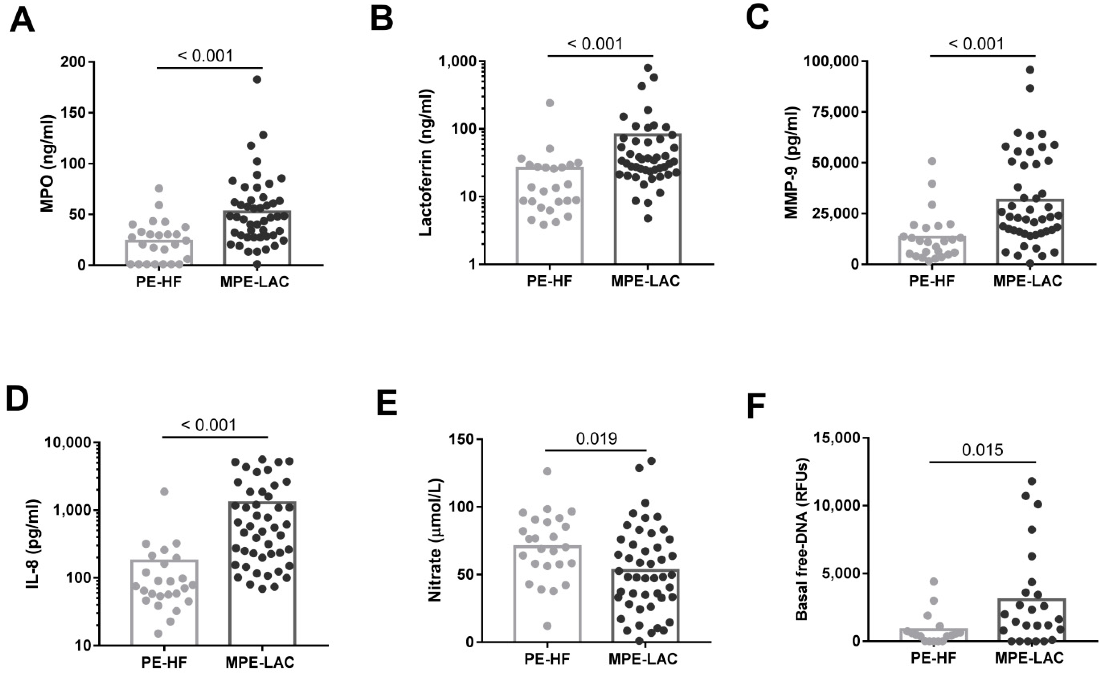
Lung Cancer Pleural Fluid Color Chart

Pleural Effusion Color Chart

Lung Cancer Pleural Fluid Color Chart

Pleural Fluid Color Chart
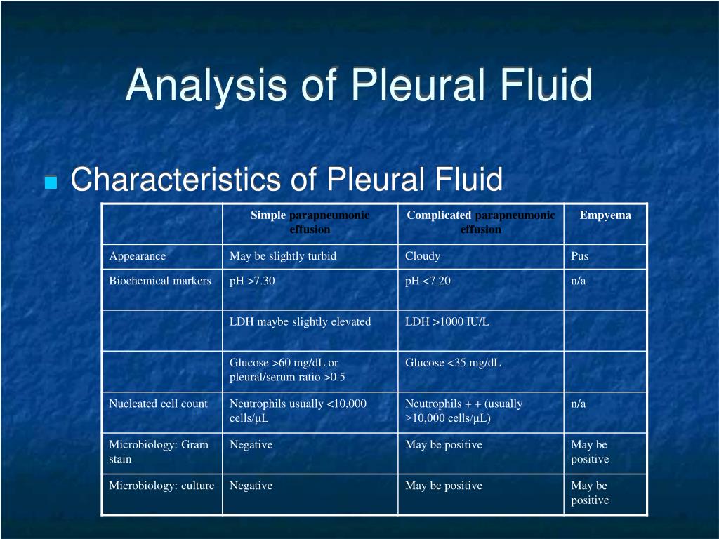
Chart Pleural Effusion Fluid Color
_CRUK_054.svg/1200px-Diagram_showing_a_build_up_of_fluid_in_the_lining_of_the_lungs_(pleural_effusion)_CRUK_054.svg.png)
Pleural effusion Wikipedia

Pleural Effusion Fluid Color
Pleural Fluid Color Chart

Pleural Effusion Color Chart
Pleural Effusion Color Chart
Your Provider Has To Get Rid Of This Fluid With Medicine Or A.
Web The Fluid Color Sign Is A Diagnostic Sign To Differentiate A Pleural Effusion From Pleural Thickening By Means Of Color Doppler Ultrasound.
Web Interpreting Pleural Fluid Results Correctly Requires An Awareness Of The Possible Aetiologies Of A Pleural Effusion And An Understanding Of The Reliability Of The Outcome Of Each.
Web Bloody Fluid Is Most Often Caused By Cancer, Pulmonary Embolus, Or Trauma;
Related Post: