Gram Positive Chart
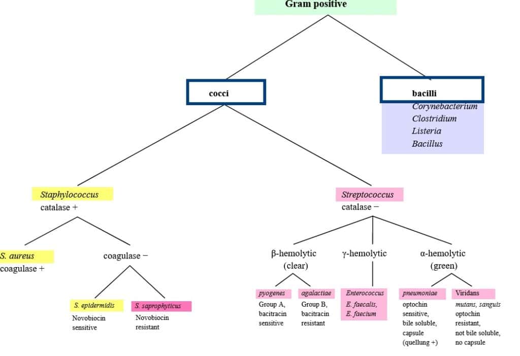
Gram Positive Chart - Actinomyces, bacillus, clostridium, corynebacterium, enterococcus, gardnerella, lactobacillus, listeria, mycoplasma, nocardia, staphylococcus, streptococcus, streptomyces ,etc. “gram positive cocci in clusters” may suggest staphylococcus species. Here’s why knowing whether the result is positive or negative is. Bacillus, clostridium, corynebacterium, listeria ,. During the gram staining process — a test that experts use to view the bacteria under a microscope — they appear purple or. Move your mouse over an item on the graphic and if your arrow turns into a hand click on it and you will go to another place in the notebook.) click on gram positives to determine how and when to perform the tests indicated above. Web gram positive bacteria types and classification. Identify similarities and differences between high g+c and low g+c bacterial groups. Web gram positive bacteria have a thick coating of peptidoglycan and stain purple with crystal violet. Learn more from the experts at uptodate. During the gram staining process — a test that experts use to view the bacteria under a microscope — they appear purple or. But, some bacteria stain either as gram positive or gram negative, depending on conditions. In a gram stain test, these organisms yield a positive result. Learn more from the experts at uptodate. The bacterial cell wall of these organisms have thick peptidoglycan layers, which take up the purple/violet stain. Web gram positive bacteria have a thick coating of peptidoglycan and stain purple with crystal violet. Move your mouse over an item on the graphic and if your arrow turns into a hand click on it and you will go to another place in the notebook.) click on gram positives to determine how and when to perform the tests indicated above. Associate various biochemical tests with their correct applications. “branching gram positive rods, modified acid fast stain positive” may suggest nocardia or streptomyces species. Gram project is a medical education resource website containing diagrams, tables and flowcharts for all your quick referencing, revision and teaching needs. Move your mouse over an item on the graphic and if your arrow turns into a hand click on it and you will go to another place in the notebook.) click on gram positives to determine how and when to perform the tests indicated above. They have thick cell walls. Gram staining works by differentiating bacteria by the chemical and. Web interpretation of key phrases. They can be found in a variety of environments and can be harmless or pathogenic to humans and other organisms. Web gram positive bacteria types and classification. The bacterial cell wall of these organisms have thick peptidoglycan layers, which take up the purple/violet stain. Web aerobic gram positive rods flowchart. “branching gram positive rods, modified acid fast stain positive” may suggest nocardia or streptomyces species. But, some bacteria stain either as gram positive or gram negative, depending on conditions. Web use flowcharts and identification charts to identify some common aerobic gram positive microorganisms. Gram positive cocci in pairs and chains may suggest streptococcus species or enterococcus species. Gram staining works. Web interpretation of key phrases. “branching gram positive rods, modified acid fast stain positive” may suggest nocardia or streptomyces species. Bacillus, clostridium, corynebacterium, listeria ,. Give an example of a bacterium of high g+c and low g+c group commonly associated with each category. Gram staining works by differentiating bacteria by the chemical and physical properties of their cell walls. Associate various biochemical tests with their correct applications. (the graphic below is clickable. Web gram positive cocci are a group of bacteria that cause various infections in humans. They have thick cell walls. They don’t retain crystal violet, so are stained red or pink with carbol fuchsin or safranin. Gram staining works by differentiating bacteria by the chemical and physical properties of their cell walls. Bacillus, clostridium, corynebacterium, listeria ,. Give an example of a bacterium of high g+c and low g+c group commonly associated with each category. “gram positive cocci in clusters” may suggest staphylococcus species. Gram negative bacteria lack this thick coating. But, some bacteria stain either as gram positive or gram negative, depending on conditions. Interpret the results of biochemical methods. Bacillus, clostridium, corynebacterium, listeria ,. The bacterial cell wall of these organisms have thick peptidoglycan layers, which take up the purple/violet stain. Gram positive cocci in pairs and chains may suggest streptococcus species or enterococcus species. They don’t retain crystal violet, so are stained red or pink with carbol fuchsin or safranin. Web interpretation of key phrases. (the graphic below is clickable. Web gram positive bacteria types and classification. “branching gram positive rods, modified acid fast stain positive” may suggest nocardia or streptomyces species. (the graphic below is clickable. Web gram positive cocci are a group of bacteria that cause various infections in humans. Bacillus, clostridium, corynebacterium, listeria ,. The bacterial cell wall of these organisms have thick peptidoglycan layers, which take up the purple/violet stain. Interpret the results of biochemical methods. Web aerobic gram positive rods flowchart. Web gram positive bacteria types and classification. Gram staining works by differentiating bacteria by the chemical and physical properties of their cell walls. They can be found in a variety of environments and can be harmless or pathogenic to humans and other organisms. Web interpretation of key phrases. “gram positive cocci in clusters” may suggest staphylococcus species. Gram positive cocci in pairs and chains may suggest streptococcus species or enterococcus species. “branching gram positive rods, modified acid fast stain positive” may suggest nocardia or streptomyces species. Interpret the results of biochemical methods. Associate various biochemical tests with their correct applications. Actinomyces, bacillus, clostridium, corynebacterium, enterococcus, gardnerella, lactobacillus, listeria, mycoplasma, nocardia, staphylococcus, streptococcus, streptomyces ,etc. The bacterial cell wall of these organisms have thick peptidoglycan layers, which take up the purple/violet stain. Give an example of a bacterium of high g+c and low g+c group commonly associated with each category. They don’t retain crystal violet, so are stained red or pink with carbol fuchsin or safranin. Web gram positive flow chart. Web gram positive bacteria have a thick coating of peptidoglycan and stain purple with crystal violet. During the gram staining process — a test that experts use to view the bacteria under a microscope — they appear purple or. Bacillus, clostridium, corynebacterium, listeria ,. Web interpretation of key phrases. Web use flowcharts and identification charts to identify some common aerobic gram positive microorganisms. (the graphic below is clickable.
Streptococcus Concise Medical Knowledge

MBBS Medicine (Humanity First) Gram positive Cocci Classification

Antibiotic Susceptibility Table for Routine Gram Positive GrepMed

Gram Positive Bacteria Flow Chart Diagram Quizlet

Microbiology Gram Positive Cocci Flow Chart Gram Positive Cocci

Gram Positive Flow Chart

Identification of Gram positive bacteria Microbiology, Medical

Gram Positive Cocci Identification Chart

Gram Positive Organisms Chart
Gram Positive Organisms Chart
Web Gram Positive Bacteria Types And Classification.
Web Aerobic Gram Positive Rods Flowchart.
The Antibiotics Listed Are Active Against E.faecalis, But Have Limited Activity For E.faecium.
In A Gram Stain Test, These Organisms Yield A Positive Result.
Related Post: